By subscribing,巴西棋牌推流博主怎么赚钱 you agree to our Terms of Use and Policies You may unsubscribe at any time.
- Imaging techniques, such as X-rays and CTs, help paleontologists and anthropologists to identify and learn about fossils and artifacts.
- Neutron imaging is a newer technology that is more effective when complex structures or soft tissue remains.
- Its distinct nuclear properties reveal hidden details, making it a powerful tool for studying fossils, artifacts, and biological specimens.
Exploring the depths of our planet's history goes beyond the expertise of paleontologists and anthropologists alone. It hinges on advanced imaging technologies that empower scientists to delve into the concealed interiors of fossils and artifacts.
From the early days of X-rays to the latest developments in X-ray CT, a new contender in the field has emerged – neutron imaging. This relatively recent technology has proven to be transformative, providing insights beyond the capabilities of traditional X-ray methods.
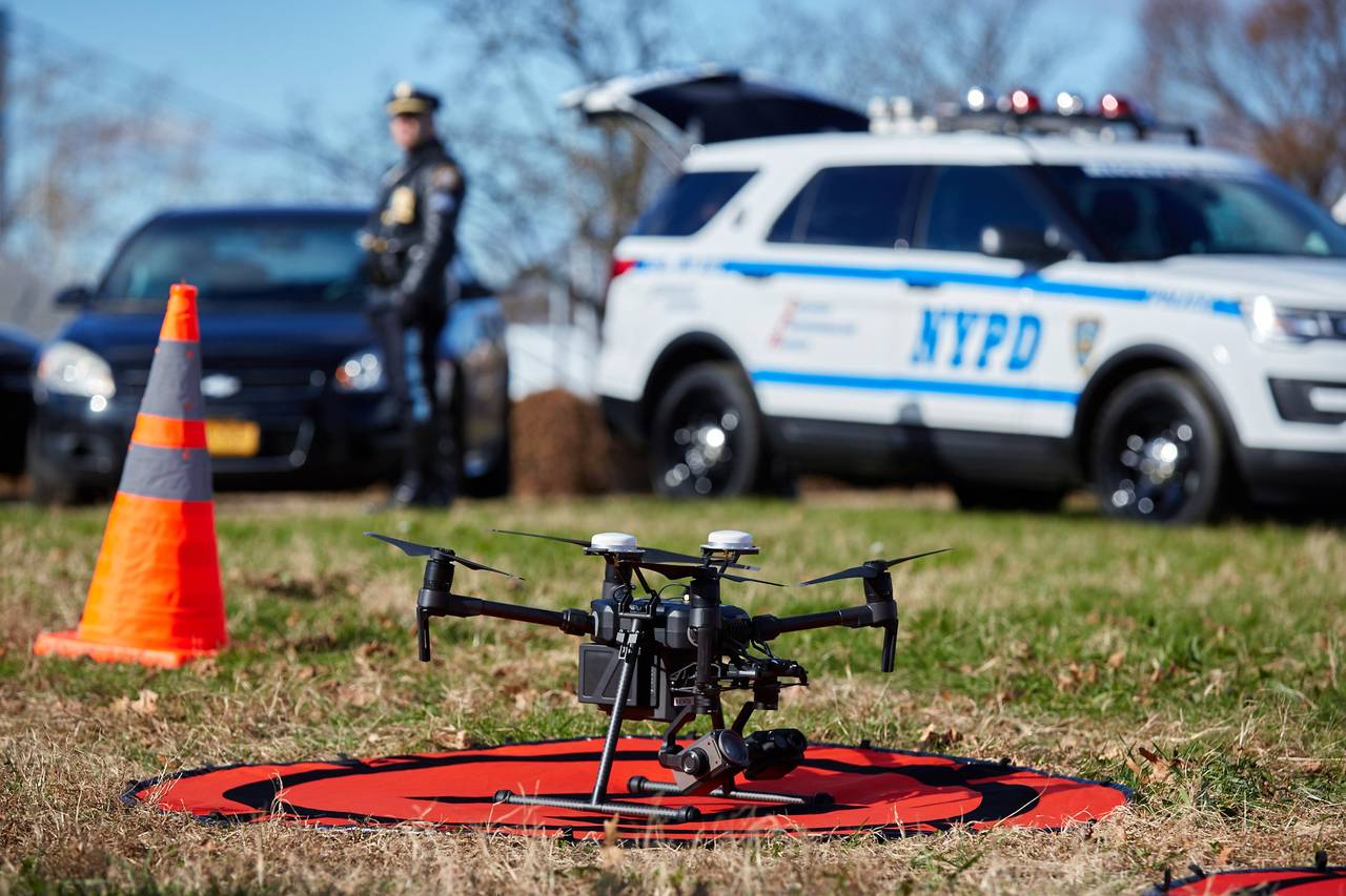
In 2022, scientists used neutron imaging to reveal the hidden secrets within Confractosuchus sauroktonos, a shattered crocodile fossil, exposing a previously unknown dinosaur species in its stomach cavity, dating back 100 million years to the Cretaceous Period.
This article explores the world of imaging techniques and how they uncover the secrets held inside fossils and old artifacts. From the cyanobacteria, the oldest known fossils dating back 3.5 billion years, to contemporary breakthroughs facilitated by imaging techniques, we unravel the secrets held within Earth's geological archives.
A brief history of imaging techniques
The origins of imaging techniques can be traced back to the unveiling of X-rays by German physicist Wilhelm Conrad Röntgen in 1895. Röntgen's discovery uncovered a new form of radiation capable of traversing human tissue but not bone or metal.
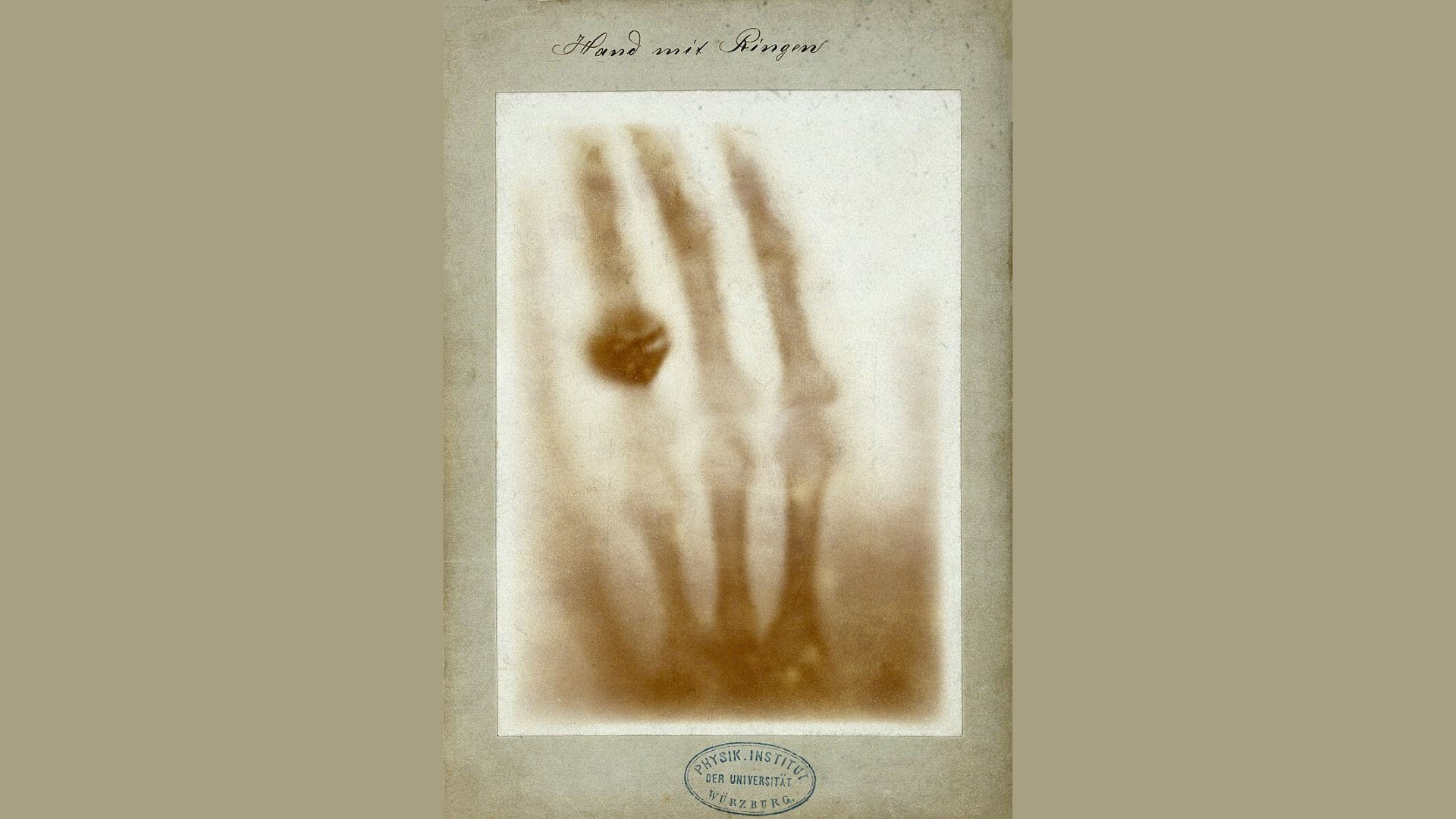
Wilhelm Röntgen
The inaugural radiograph, capturing Röntgen's wife's hand, marked the first-ever photograph of a human body part using X-rays—the use of X-rays in paleontology dates back to the early days of its discovery in 1896.
Pioneering attempts by Brühl in Berlin and Lemoine in Paris within just nine months of Röntgen's initial discovery set the stage. Unfortunately, the physical evidence from these early efforts has been lost.
Substantive work on fossil imaging began in 1906. However, it was W. M. Lehmann who, from 1934 onwards, systematically and widely employed X-ray techniques in the investigation of Hunsrück slate fossils. Notably, Wilhelm Stürmer is regarded as the father of modern techniques for studying Hunsrück slate fossils. During the 1970s, he successfully captured images of soft-bodied organisms and internal organs, pioneering a mobile X-ray laboratory at the discovery site.
The history of X-ray computed tomography (CT) reaches back to at least 1917, rooted in the mathematical theory of the Radon transform.
In 1984, a significant leap occurred with the application of CT scanning to a mammalian cranial fossil by paleoanthropologist Glen Conroy and radiologist Vannier. High-resolution CT scans revealed density differences among cranial cavities, air spaces, and bone, marking a milestone in the intersection of paleontology and advanced imaging technology.
Today, X-ray CT is the go-to imaging technique for studying fossils and old artifacts. Its non-destructive nature allows for thoroughly examining fossils without compromising their integrity.
X-ray CT's ability to create detailed 3D models has revolutionized how researchers analyze and interpret fossilized remains. This technology not only aids in identifying species but also offers a deeper understanding of anatomical structures and evolutionary processes.
Neutron imaging
Imaging techniques have come a long way since the discovery of X-rays in 1895. X-rays function by penetrating an object, where denser materials absorb more radiation, presenting as darker regions in the resultant image.
For example, human tissue, being less dense, allows more X-rays to pass through, resulting in lighter areas on the X-ray image. In contrast, denser structures like bones absorb more X-rays, appearing as darker areas. This differential absorption enables the visualization of internal structures.
One of the challenges is differentiating materials with similar densities. This limitation can make it challenging to understand the entire composition and intricacies of certain fossils or artifacts, particularly those with complex internal structures or soft tissue remains.
On the other hand, neutron imaging utilizes neutron beams. Neutrons are electrically neutral particles, interacting primarily with an atom's nucleus through processes like scattering.
Unlike X-rays, which can be influenced by electron density, neutrons penetrate materials without significant interaction with electrons. This difference makes neutron imaging particularly effective for materials having low electron density but distinct nuclear properties.
Additionally, certain elements, like hydrogen, stand out more in neutron imaging compared to X-rays. This property is valuable in studying organic materials, making neutron imaging a powerful tool for investigating biological specimens, archaeological artifacts, and other materials where traditional X-ray methods might fall short.
The history of neutron imaging can be traced back to James Chadwick's discovery of neutrons in 1932. Hartmut Kallmann and E. Kuhn demonstrated neutron radiography in the late 1930s, revealing materials emitting radiation under neutron bombardment.

National Institute of Standards and Technology
Notably, reasonable-quality neutron radiographs emerged in 1955, pioneered by J. Thewlis in the UK, following Peters' initial low-quality attempts in 1946. However, until recently, neutron imaging's potential for exploring fossils and old artifacts was proven, thanks to the NeXT facility at the National Institute of Standards and Technology in Gaithersburg, led by physicist Jacob LaManna.
A dinosaur-eating crocodile
The crocodile neutron imaging story involves the discovery of Confractosuchus sauroktonos, a shattered crocodile fossil found in Australia. Initially, X-ray CT scans were used to create a 3D map of the interior, aiming to isolate individual bones and reconstruct the crocodile virtually. However, challenges arose due to iron-rich stone surrounding the bones, hindering clear X-ray images.
The researchers opted for neutron imaging at the Australian Centre for Neutron Scattering to overcome this obstacle. Chemist Joseph Bevitt, who specializes in neutron imaging, scanned the fossil using subatomic neutron particles.
The neutron scans not only revealed the expected crocodile bones but also unveiled a surprising discovery—a dinosaur leg bone within the crocodile's stomach cavity. This unexpected find led to the identification of a previously unknown species of dinosaur, providing valuable insights into the ancient ecosystem.
This finding has significant implications for understanding the ancient ecosystem and predator-prey relationships during the Cretaceous Period, approximately 100 million years ago.
The discovery of the dinosaur species inside the crocodile's stomach, showing bite marks and being broken into pieces, provides a clear insight into ancient interactions.
The neutron imaging not only showcased the power of this technology in revealing intricate details in challenging conditions but also demonstrated its role in unearthing unexpected and valuable information about long-extinct species and their ecological dynamics.
Shang dynasty’s dagger-axes
The crocodile case study demonstrates the efficacy of neutron imaging in fossil studies. However, challenges arise in cases like the unsuccessful investigation of 3,000-year-old dagger-axes from China's Shang dynasty. These ceremonial weapons, featuring jade blades and bronze handles, posed difficulties due to their diverse composition.
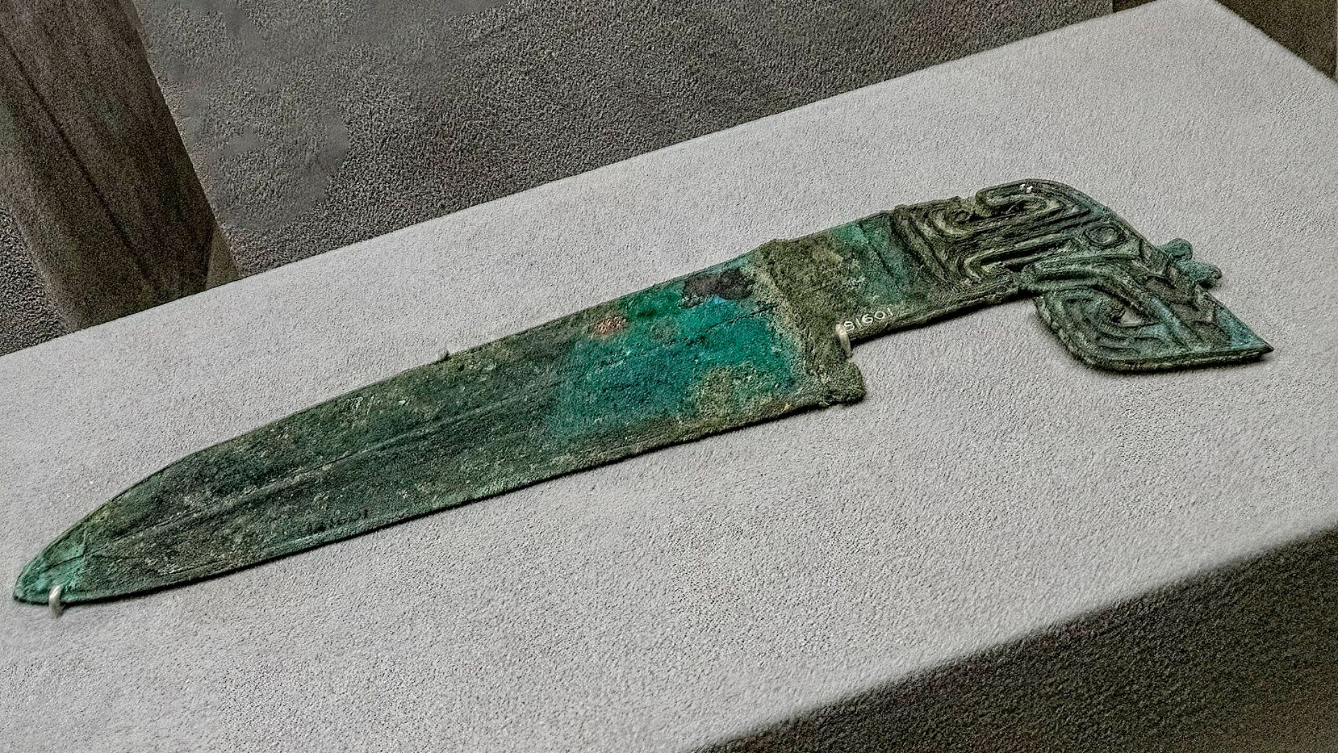
Mary Harrsch
Initially, there was a contemplation of utilizing neutron imaging to study the dagger. However, there was a significant drawback: neutron beams used in imaging can make objects radioactive, posing potential risks and uncertainties about the level and duration of radioactivity induced in the samples.
The conservator at the Smithsonian's Freer Gallery of Art, Ariel O'Connor, sought to test the feasibility of neutron imaging without risking the precious artifacts. A replica was created and exposed to X-ray and neutron scans without risking authentic artifacts.
While the X-ray struggled with brass components, the neutron scan revealed crucial details without inducing radioactivity in the replica. Despite success with the test, conservators expressed reluctance to use neutron imaging on genuine artifacts due to uncertainties and potential alterations.
This case underscores neutron imaging's dual nature—unveiling details while posing risks, especially with culturally significant artifacts. Balancing scientific exploration with historical preservation is crucial in such delicate scenarios.
Navigating scientific exploration and cultural artifact preservation in an evolving field
The utilization of imaging technologies, from traditional X-rays to the more recent neutron imaging, has significantly advanced our understanding of fossils and artifacts.
While neutron imaging has advantages, striking a balance between scientific exploration and the preservation of cultural artifacts remains a critical consideration in this evolving field.
Neutron imaging is still a new technology and requires refinement to address challenges such as potential radioactivity associated with certain materials. As this technology matures, it promises further to enrich our exploration of Earth's geological archives, providing deeper insights into the intricate stories embedded within fossils and artifacts.